Viewzyvex
Your Beginner Guide to Identifying Common Minerals
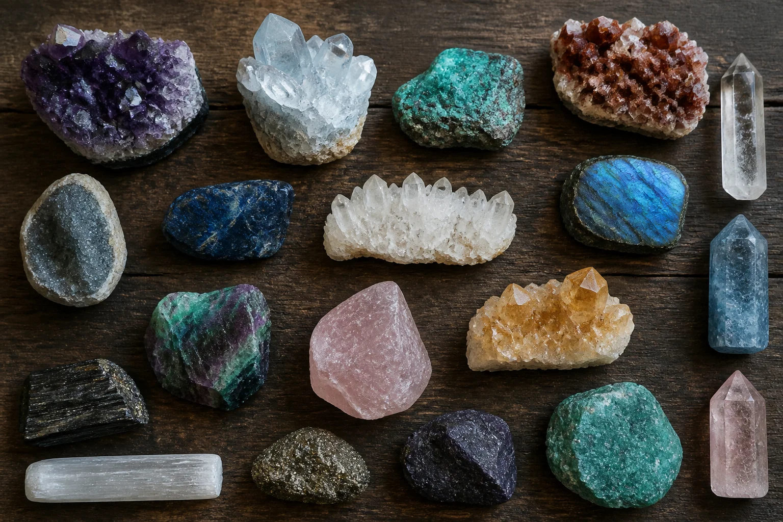
Picking up an unusual stone from a riverbed or purchasing a dusty box of specimens sparks an immediate curiosity. Unlocking the identity of that find requires looking past the initial colors and shiny surfaces.
Geology relies on systematic testing rather than guesswork. Minerals possess hidden signatures written in their atomic structures, and simple physical tests reveal those secrets on a workbench.
The resistance of a mineral surface to scratching forms the backbone of basic identification. Friedrich Mohs devised a scale ranging from one to ten, where talc sits at the softest end and diamond crowns the summit.
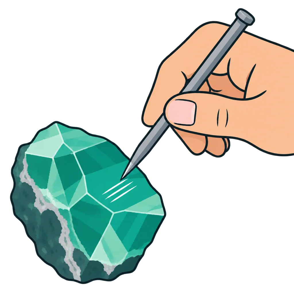
Testing hardness requires everyday objects with known resistance values rather than specialized laboratory tools. A fingernail scratches gypsum easily, while a steel pocket knife leaves a mark on fluorite but slides harmlessly across quartz.
Pressing too hard or testing weathered outer crusts produces misleading results. Always scrape away the oxidized surface layer and test on a fresh fracture beneath it.
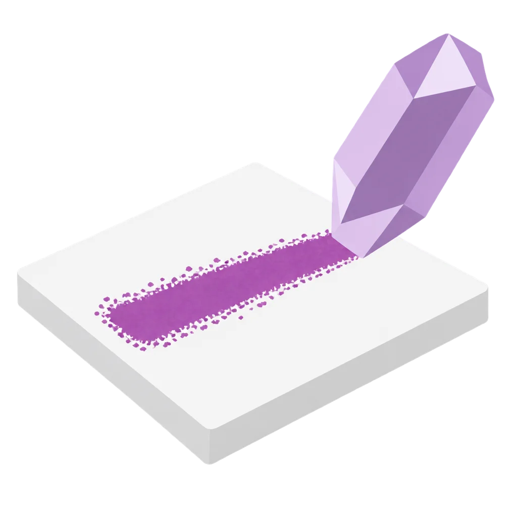
The external color of a mineral often lies, but the powder it leaves behind tells the absolute truth. Dragging a specimen across an unglazed porcelain plate produces a fine streak of powdered mineral.
Pyrite fools many beginners with its brilliant golden metallic flash, leaving a dense black streak across the plate instead of gold. Hematite shifts from metallic gray lumps to a distinct reddish-brown powdery line.
Light interacts with crystal surfaces in distinct ways that define luster categories. Vitreous luster resembles ordinary window glass, while pearly surfaces catch light with a soft, milky sheen.
Metallic specimens reflect light like polished chrome or raw iron bars, casting sharp shadows under a desk lamp. Greasy or resinous textures make the stone look coated in oil or amber.
Observing luster requires turning the sample slowly beneath a single light source. Direct sunlight washes out subtle shifts, so a directional flashlight works best for catching surface reflections.
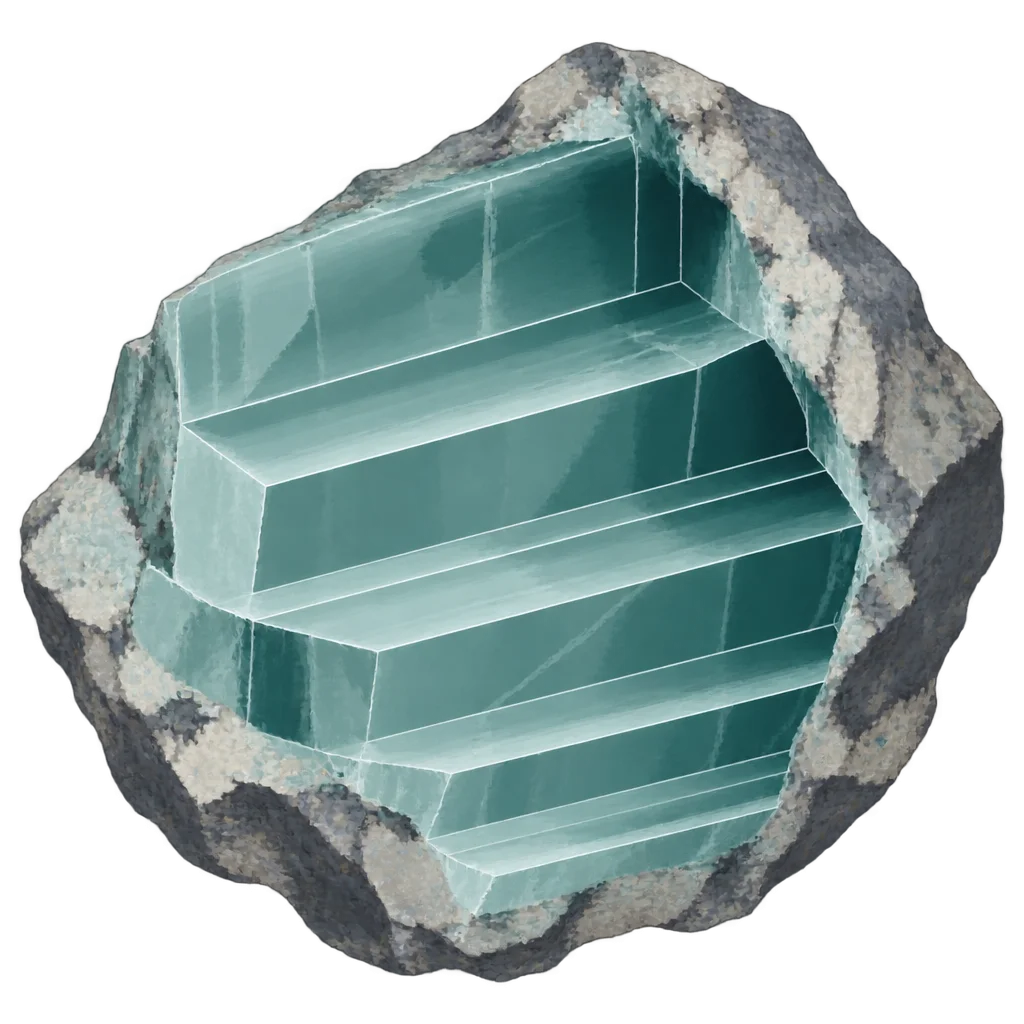
Internal atomic bonds create lines of weakness where minerals prefer to break smoothly. Counting distinct cleavage directions and measuring the angles between them separates mica sheets from cubic galena.
Mica peels apart into flexible elastic sheets because bonds remain exceptionally weak in one single direction. Calcite splits into tilted rhombs with precise angles, reflecting light across flat, geometric faces rather than jagged fractures.
Conchoidal fracture differs entirely by producing curved, shell-like ripples across glass-like rocks. Quartz breaks this way, ignoring flat planes completely and creating sharp, razor-thin edges.
Amateurs constantly confuse clear quartz crystals with transparent calcite rhombs on flea market tables. Simple physical tests separate these two common minerals without damaging either specimen.
Dropping a tiny splash of vinegar onto the stone settles the debate instantly. Calcite fizzes quietly as acid releases carbon dioxide bubbles, while quartz remains completely unbothered by household acids.
Scratching the surface provides a second backup test for verification. Quartz rates a seven on the hardness scale and bites deeply into ordinary glass, whereas calcite scratches at a three and yields to a copper coin.
Building confidence with minerals takes patience, a handful of basic tools, and a willingness to look closely at ordinary stones. Every scratch and streak brings clarity to an otherwise silent collection.

Jan de Jong
Lead Mineralogist
Sanne Bakker
Field Collector
Lieke de Vries
Lab Technician
Maarten van Dijk
Curator

Lead Mineralogist

Field Collector

Lab Technician

Curator